
Panoramic Dental X-Ray in Montreal: What Does It Show?
A panoramic dental X-ray, also known as a
Panorex, panoramic radiograph, or OPG,
provides a broad view of the teeth, jaws, and several surrounding structures
in a single image.
It is particularly useful for evaluating wisdom teeth, impacted teeth,
the jawbones, certain infections, and different abnormalities.
However, it does not replace the clinical examination
or smaller intraoral X-rays when more detailed images are required.
At Dentistes sur Drummond in downtown Montreal,
dental X-rays are recommended according to each patient’s individual needs,
symptoms, history, clinical examination,
and previously available images.
The key point:
a panoramic X-ray provides an excellent overall view,
but it does not show every individual tooth with the same level of detail
as an X-ray taken inside the mouth.
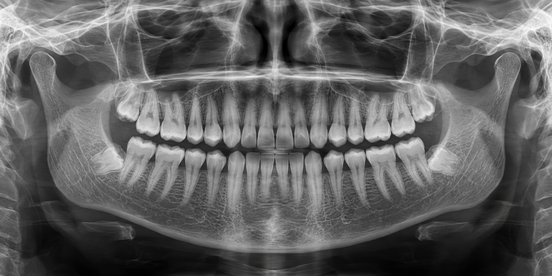
of the teeth and jaws. This image is presented for general information
and must not be used for diagnostic purposes.
What Is a Panoramic Dental X-Ray?
A panoramic radiograph is a two-dimensional dental image.
Unlike smaller dental X-rays,
no sensor is placed at the back of the mouth.
During the exposure, the machine rotates around the patient’s head.
It creates an overall image that may include:
- the upper and lower teeth;
- the dental roots;
- the upper and lower jawbones;
- teeth that have not yet erupted;
- wisdom teeth;
- portions of the maxillary sinuses;
- portions of the jaw joints;
- several anatomical structures surrounding the teeth.
Because the jaws are curved and the final image is flat,
some structures may appear slightly enlarged,
distorted, or less sharply defined.
Why Might a Dentist Recommend a Panoramic X-Ray?
A panoramic radiograph may be recommended when a broad view
of the teeth and jaws is needed to establish a diagnosis
or plan treatment.
It may be useful for:
- evaluating the position of wisdom teeth;
- locating impacted or unerupted teeth;
- assessing dental growth and development;
- examining the jaws following trauma;
- planning certain dental extractions;
- obtaining an overview before orthodontic treatment;
- evaluating areas where teeth are missing;
- contributing to the planning of certain implant or surgical procedures;
- investigating certain jawbone lesions or abnormalities;
- evaluating unexplained pain or swelling.
A panoramic X-ray is not automatically required
simply because someone is a new patient.
It is prescribed when it is expected to provide information
that is useful for diagnosis or treatment.
Why Is It Useful for Wisdom Teeth?
A panoramic radiograph allows the dentist to view all four areas
where wisdom teeth usually develop in a single image.
It can help evaluate:
- whether the wisdom teeth are present;
- whether they are erupted, partially erupted, or impacted;
- their direction and angulation;
- the amount of available space;
- their relationship with neighbouring teeth;
- possible abnormalities surrounding a tooth.
In the lower jaw, it also provides an initial indication
of the relationship between the wisdom-tooth roots
and the mandibular canal, which contains an important nerve.
When this relationship appears very close
or more precise three-dimensional information is needed,
a CBCT scan may be recommended.
Can a Panoramic X-Ray Show Cavities?
A panoramic image may sometimes reveal relatively large cavities.
However, it is generally not the most precise image
for detecting small cavities between teeth
or underneath certain fillings and crowns.
To evaluate decay between the teeth,
the dentist may recommend bite-wing X-rays.
A patient may therefore need both a panoramic radiograph
and a few smaller intraoral images.
These images are not necessarily duplicates:
they provide different and complementary information.
What Can a Panoramic X-Ray Reveal?
Depending on the situation, it may help the dentist observe:
- the presence and general position of the teeth;
- impacted teeth;
- missing teeth;
- certain significant infections;
- advanced bone loss;
- certain jaw lesions or abnormalities;
- certain fractures;
- the general shape of the jaws;
- portions of the maxillary sinuses;
- portions of the temporomandibular joints.
An abnormality visible on a panoramic image
does not always provide a final diagnosis.
More focused X-rays, CBCT imaging,
specialist consultation, or other tests
may sometimes be required.
What Are Its Limitations?
A panoramic radiograph covers a large region,
but it provides less fine detail than certain intraoral X-rays.
It may not be sufficient for:
- detecting very small cavities between teeth;
- evaluating a small infection around a root;
- examining the fine details of root canal treatment;
- measuring every dimension of the jawbone precisely;
- evaluating the soft tissues of the mouth;
- diagnosing a jaw-joint condition by itself;
- replacing the dentist’s clinical examination.
It also does not replace examination of the tongue,
cheeks, palate, and other oral tissues.
Many soft-tissue conditions cannot be seen
on a panoramic X-ray.
What Is the Difference Between a Panoramic X-Ray and CBCT?
A panoramic X-ray produces an overall image in
two dimensions.
It is appropriate for many common assessments.
A CBCT scan, or cone-beam computed tomography,
produces three-dimensional images.
It can display structures from different angles
and provide more precise information about bone,
roots, nerve canals, and complex anatomical areas.
CBCT is not automatically preferable.
It is recommended when three-dimensional information
can influence the diagnosis or treatment plan.
What Happens During a Panoramic X-Ray?
The procedure is quick, painless,
and usually requires no special preparation.
-
You will be asked to remove your glasses,
earrings, necklaces, jewellery, or other metal objects. - You will stand or sit in the machine.
- You will bite gently on a small positioning guide.
- Your head will be aligned using supports and positioning lights.
-
You will be asked to keep your tongue against the roof of your mouth
and remain completely still. - The machine will rotate around your head for a few seconds.
Because no large sensor is placed at the back of the mouth,
panoramic imaging may be more comfortable
for patients with a strong gag reflex.
Why Is Correct Positioning Important?
The quality of a panoramic radiograph depends greatly
on patient positioning.
If the head is too far forward, too far back, tilted, or rotated,
some teeth may appear blurred, widened, or narrowed.
Movement during the exposure may also reduce image quality.
Following the dental team’s instructions
helps produce a clear image on the first attempt
and reduces the possibility that it will need to be repeated.
Is a Panoramic X-Ray Safe?
Like all dental radiographs,
a panoramic X-ray uses X-radiation.
It is recommended when the expected information
provides a clinical benefit to the patient.
Digital technology, appropriate positioning,
and suitable exposure settings help obtain
the required information while limiting exposure.
Dental X-rays are not taken according to the same schedule
for every patient.
The dentist considers:
- the patient’s age;
- symptoms;
- current oral health;
- risk factors;
- the treatment being considered;
- previously available X-rays.
What If I Am Pregnant or May Be Pregnant?
Always tell the dental team if you are pregnant
or if there is a possibility that you may be pregnant.
The dentist will consider the reason for the image
and the urgency of the situation.
A non-urgent image may sometimes be postponed,
while an X-ray needed to diagnose pain,
infection, or another important condition
may be taken using the appropriate protocols.
I Already Have a Recent Panoramic X-Ray. Must It Be Repeated?
Not necessarily.
A recent panoramic image from another dental clinic
can often be used when it is clear,
complete, and of diagnostic quality.
We recommend having it transferred before your appointment.
The dentist will consider:
- the date of the radiograph;
- its quality and clarity;
- the areas that are visible;
- possible changes since it was taken;
- the reason for your current consultation.
A new image may be needed when the previous radiograph
is too old, incomplete, blurred,
or does not answer the current clinical question.
Important information about fees:
providing a recent and usable panoramic X-ray
may prevent the same image from being repeated,
but it does not replace the clinical examination
and does not reduce the examination fee.
Is the Panoramic X-Ray Included in the Examination Fee?
At Dentistes sur Drummond,
the panoramic X-ray is not included
in the examination fee.
When it is clinically indicated and taken at our clinic,
it is a separate service
and is billed separately.
The examination fee remains the same
when a patient provides a recent panoramic image,
because the dentist must still perform the clinical examination,
interpret the image, establish an independent diagnosis,
and explain the recommendations.
Frequently Asked Questions
Do I Need a Panoramic X-Ray Every Year?
No. There is no single interval that applies to every patient.
A new panoramic image is recommended
when it can provide relevant information
for diagnosis or treatment.
Does a Panoramic X-Ray Hurt?
No. The machine does not touch your teeth,
and the exposure takes only a few seconds.
Does It Replace All Smaller Dental X-Rays?
No. A panoramic image provides an overall view,
while intraoral X-rays show individual teeth
in greater detail.
Does It Show Every Cavity?
No. It may reveal certain larger cavities,
but small cavities between the teeth
may require bite-wing X-rays.
Can It Diagnose Oral Cancer?
It may sometimes reveal an abnormality within the jawbone,
but it does not replace a clinical examination
of the mouth’s soft tissues.
Any suspicious finding must be evaluated
and may require additional testing.
Why Must I Remove My Jewellery?
Metal objects can create artefacts
that hide portions of the teeth or jaws.
Removing them helps produce a clearer image.
Panoramic Dental X-Rays in Downtown Montreal
At Dentistes sur Drummond,
we have an on-site digital imaging system
that allows us to take a panoramic dental X-ray
when it is clinically indicated.
Each image is interpreted together with your clinical examination,
symptoms, history, and other available information.
Our goal is to obtain the images that are useful
for your situation without taking unnecessary X-rays.
Dentistes sur Drummond
1227 Drummond Street
Montreal, Quebec H3G 1V8
Telephone:
514-939-2492
You can also visit our page about our
dental technologies
.
This article provides general information.
The need for and type of dental imaging recommended
will vary according to each patient’s clinical situation.
Image Reference
Main image — Illustration of a panoramic dental X-ray
Original image generated for this Dentistes sur Drummond article.
This image is an illustration and is not an X-ray from a real patient.
It must not be used for diagnostic purposes.
Informational References
-
American Dental Association — X-Rays/Radiographs:
View the reference
-
RadiologyInfo.org — Panoramic Dental X-Ray:
View the reference


